Why Fish Should Be On Your Diet List?

Fish, like salmon, mackerel and albacore tuna, that high in omega-3 fatty acid is good for the heart because omega-3 fatty acid could reduce the risk of coronary heart disease. Perhaps this is why American Heart Association (AHA) advice people to eat at least 2 servings of fish a week.

Despite concerns on the impact of increased exposure to mercury in fish, Swedish researchers from Umea University in Sweden argued in their paper published in November 2010 in ‘American Journal of Clinical Nutrition’ that the benefits of consuming fish still outweigh the harmful effects of mercury. However, they also advised people to avoid eating fish high in mercury, which include perch, shark, swordfish and halibut.

More than 900 Swedish men and women took part in a study that required them to answer questionnaires about the amount of fish in their diet. Their mercury and selenium levels in red blood cells were analyzed.

While the mercury levels were generally low for Scandinavians, participants who had higher mercury level in their red blood did not have a higher risk of cardiac problems. It is possible that the protective nutrients in fish offset any harmful effect of mercury at these low levels of mercury, as explained by the researchers.

Meanwhile, the researchers admitted that relying on the participants’ memories on the amount of fish consumed could have drawbacks as this could have an impact on the results obtained. They also highlighted one finding from their study: participants who had elevated traces of selenium in their red blood cells appeared to have higher risk of sudden cardiac death.

Selenium is a trace mineral that is essential to good health but only small amounts are needed. It appears to act as an antioxidant that can reduce risk of cardiovascular disease and cancer.

Nevertheless, previous findings on effect of selenium on heart were mixed. Several studies suggested risk of heart disease and stroke might be reduced by about 40 percent by an added 100 mg per day of dietary selenium. On the other hand, a study by researchers from University of Warwick found that total cholesterol levels increased by 8 percent with an increase of 10 percent in LDL (so called bad cholesterol) in participants having more than 1.2 mol/L of selenium in their blood, leading to increase in risk of cardiovascular disease.

As such, the Swedish researchers felt that the health effect of selenium should further be probed.

Weight Loss Could Help Ward Off Diabetes!

Insulin resistance sometimes combined with absolute insulin deficiency cause Type-2 diabetes to develop. Insulin resistance is a condition in which cells cannot use insulin properly. Type-1 and gestational diabetes are the other two main types of diabetes.

Of the many risk factors identified for Type-2 diabetes, obesity, sedentary lifestyle and unhealthy eating habits are somewhat associated with weight gain. High cholesterol, high blood pressure, family history and genetics, and increased age can also raise the likelihood of getting Type-2 diabetes.

A study conducted by researchers from Johns Hopkins University in Baltimore found that Type-2 diabetes could possibly be reversed if obese diabetics go for a weight-loss surgery. Their findings published on August 16, 2010 in the medical journal, ‘Archives of Surgery’ indicated that 75 percent of obese diabetics who had undergone weight-loss surgery (bariatric surgery) could actually stop taking diabetes medications within 6 months of their operation.

Researchers carried out one of the few studies examining how surgery can affect health cost in Type-2 diabetics by analyzing insurance claims data from 2,235 patients who underwent bariatric surgery during a 4-year period.

The results showed that among the diabetic patients who had bariatric surgery, only one quarter were still taking diabetes drugs 6 months later. In fact, the number kept falling: 12 months after the surgery, less than 20 percent of patients were taking medications and 24 months after surgery, only 15 percent were still been prescribed with medications.

It has long been known that diabetes and obesity can rarely be reversed once they are developed. This is definitely good news for obese Type-2 diabetics since the need for chronic medications can be eliminated and the overall medical cost could be reduced too. More importantly, this can greatly reduce the risk of developing heart considering the fact that diabetes is a risk factor for heart disease.

Nevertheless, it seems that bariatric surgery would be the only solution that most of the obese diabetics can opt for in order to substantially reverse both obesity and Type-2 diabetes, at least for the time being until experts can find a successful non-surgical methods.

Liver - Development

In the fetus, the liver develops from the hepatic diverticulum, and draw blood from the arteries that carry blood from the vitelline yolk sac. Top diverticulum causes hepatocytes and bile ducts, the lower the gallbladder with cystic duct.

During fetal development, the main source of blood to the liver is the umbilical vein that transport nutrients to the fetus. The umbilical vein enters the abdomen at the navel and moves next to the free end of the falciform muscle on the bottom exterior of the liver, where it joins the left branch of portal vein. Ductal Aranza brings blood from the left branch of portal vein branches of the left hepatic vein and then into the lower vein vein so that blood from the placenta to bypass the delivery of the fetus.

Liver in the fetus develops during pregnancy and do not perform the normal function of blood purification. The activities associated with digestion, because the fetus is fed directly from the mother's bloodstream through the placenta. Releases fetal liver stem cells migrate to the thymus and fetal red blood cells and T cells after birth, the shift of stem cells in bone marrow.

Within five days after birth, the umbilical vein and ductus venosus closes first, Teresa and the ligamentum venosus ligament seconds. In the case of cash or portal vein hypertension, the umbilical vein again.

Liver Diseases

Liver Diseases

Nmerous liver diseases are accompany by jaundice caused by augmented levels of bilirubin in the body. Bilirubin is the result of degradation of hemoglobin of dead red blood cells are normally removed by the liver and excreted in bile.

• "With hepatitis, inflammation of the liver, is caused by different viruses, but also some toxic substances, autoimmune diseases and inherited conditions;

Liver cirrhosis is the formation of fibrous tissue in the liver to kill hepatocytes, respectively. Liver cell death caused by viral hepatitis, alcohol poisoning or other toxic substances;

• With Hemochromatosis is an inherited disorder that causes iron accumulation in the body, leading to long-term liver damage;

• Benign tumors such as adenoma, "angioma" focal nodular hyperplasia.
liver cancer as the primary tumor or carcinoma cholangiocarcinoma, or metastasis of cancer to other parts of the digestive system;

Wilson disease is an inherited disorder that causes copper accumulation in the body;

Primary sclerosing cholangitis, an inflammatory autoimmune disease carries bile;
bilirubin metabolism

Pime biliary cirrhosis, autoimmune disease of minute rage ducts;

Budd-Chiari syndrome or hepatic vein obstruction;

Gilbert's syndrome, a genetic disorder of bilirubin metabolism.

There are also many pediatric liver disease. proper liver function can be verified by a number of specialized clinical studies, which measure the presence or absence of typical enzymes, metabolites, or substances associated with the regular activities of the

liver diseases

.

Liver - Physiology

Liver Physiology

Liver functions were performed liver cells to hepatocytes.
• The liver produces and secretes bile, used to emulsify fats. Part of the bile is poured directly into the duodenum, the part stored in the gallbladder.
• liver performs many functions in carbohydrate metabolism:
• gluconeogenesis or synthesis of glucose from certain amino acids, lactic acid or glycerine;
• glycogenolysis and the formation of glucose from glycogen (also occurs in the muscles);
• glycogen, or glycogen from glucose;
• destruction of insulin and other hormones;
• protein metabolism.
• Liver is also involved in lipid metabolism:
• There is a synthesis of cholesterol;
• the synthesis of triglycerides.
• hemoglobin, the liver breaks', creating metabolites that are added to bile as pigment.
• liver breaks highly toxic substances and various drugs in a development called drug metabolism. This process can lead to poisoning, when the metabolite is more toxic than its predecessor.

liver physiology
• The liver converts ammonia "in the bridge.
• liver serves as a repository for numerous substances, as well as glucose (as glycogen), vitamin B 12, copper and also iron.
• In the fetus until the third month, the liver is the major site of production of red blood cells are replaced in this capacity from bone marrow at 32 weeks of pregnancy.
• liver reticuloendothelial system contains highly specialized cells of the immune system as "filters" against anti carried by the portal vein system.

Currently there is no artificial organ able to emulate all the functions of the liver. Some of them are emulated by liver dialysis , an experimental treatment for severe cases of liver failure.

Detoxification
One of the main activities of the liver is the detoxification of the body of toxins, wastes and other harmful elements. The most important of these shares Detoxifying is the transformation of ' ammonia present in the blood (toxic substance derived from proteins ) in a substance tolerable at higher concentrations, the ' urea . The urea is then pumped back into the blood.

Relations with other bodies
The cytologic features of hepatocytes reflect the detoxifying function of the gland. They already have an endoplasmic reticulum smooth high abundance. Quest'organulo fact, in addition to chairing the synthesis of some steroids is intensely involved in the activity of its detossifacazione of potentially harmful or foreign molecules (alcohol, drugs, etc.).

Liver Cells

Liver Cells

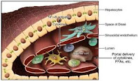
The four major cell types that are found in the liver are hepatocytes, stellate cells, sinusoidal endothelial cells and Kupffer cells.

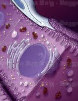
Hepatocytes are the most numerous cells of the liver, represent 80% of the volume and about 60% by number. Their shape is multifaceted, with a number of areas ranging from six to twelve, their diameter ranges from 20 to 30 microns. They are often multinucleated and tetraploid, with the number of cores that can be up to four, a large nucleolus, rough and smooth endoplasmic reticulum well developed, numerous cisternae of Golgi, ribosomes, lysosomes, mitochondria, peroxisomes, which are both one of the cell types in which the organelles are more developed and presented, due to the high metabolic needs and the wide variety of tasks which they perform.
Liver Cells

In a well-nourished body is not difficult to detect moderate amounts of glycogen and lipid vacuoles, or in the case of an overdose of iron, vacuoles or aggregates of ferritin and hemosiderin. The cytoplasm is eosinophilic background for the large number of mitochondria but with numerous basophilic granules due to the rough endoplasmic reticulum and ribosomes. You can find lipofuscin granules of golden-brown. The pole is fitted with sine dell epatocita many long and irregular microvilli on average 0.5 microns, the surface area alone is equal to 2 / 3 of the entire cell. Two adjacent hepatocytes form their plasma membranes with the bile canaliculi and are joined by tight junctions to prevent the entry of bile into the interstices, in the rest of the cell are the most common desmosomes and gap junctions. At the level of bile canaliculi accumulating numerous exocytosis of vesicles containing precisely to secrete bile into canaliculi.
stellate cells

Liver Cells

Ito or stellate cells, mesenchymal and far fewer of hepatocytes, are placed between the plates at the base of hepatocytes, and have a star-shaped or irregular. Their cytoplasm is rich in lipid vesicles containing vitamin A, and their job is to secrete the main constituent materials of the matrix, including type III collagen and reticulin. They are essential in liver regeneration following injury or surgery because they secrete growth factors responsible for good regeneration capacity of the liver. In case of injury can replace damaged hepatocytes and by the secretion of collagen and other structural proteins, forming scar tissue from the area 3 of each berry. Other substances secreted from their body contribute to homeostasis.
Sinusoidal endothelial cells

Liver Cells

Sinusoidal endothelial cells are fenestrated endothelium of venous sinusoids of the liver. They have flattened, with an oval centrally located, scant cytoplasm containing numerous vesicles transcitotiche, are joined by adherens junctions. The perforation between these cells are very large and combined in complex with an average diameter of 100 microns, so that blood can easily spill over the space of Disse and come into contact with the microvilli of hepatocytes.
Kupffer cells

Liver Cells

Kupffer cells, macrophages of the liver, are derived monocytes, they are in the lumen of the venous sinusoids. Their shape is variable and irregular, with numerous protrusions typical of cells of the macrophage that extend into the lumen of the sinusoid. Their function is to remove any debris by phagocytosis in the blood flow in the liver, but may also stimulate the immune system through the secretion of numerous factors and cytokines. Remove aged or damaged red blood cells acting as a complement to the spleen (which may be substituted in case of splenectomy).Liver Cells

Liver - Microscopic Anatomy

General structure
The liver is covered (except for bare triangular area on the upper surface) from the visceral peritoneum, consisting of the mesothelium, a single layer of surface cells and underlying tissue extraperitoneal. It is also completely surrounded by loose connective tissue component of the capsule of Glisson, who is also the neurovascular bundle at the hilum. Out from it and trabecular connective tissue septa that penetrate the liver parenchyma, dividing it in the portal tracts. Within each area there is a branch of the portal vein, a hepatic artery, a bile duct (portal triad) and often also small lymphatic vessels and nerve branches.

The parenchyma is composed of plates instead of the complex three-dimensional structure and consist of a single layer of cells, or hepatocytes, the major liver and those that perform almost all of its metabolic functions. Each layer of hepatocytes is separated from each other by a venous sinusoid, resulting from the branching of the portal vein which runs in the portal space. It takes up space portal to centrilobular vein. Hepatocytes do not adhere to the venous sinusoids, but are separated by a small intercellular space dilation in pathological conditions (0.2-0.5 μm), said space of Disse.

In the space of Disse contains mainly fibers of collagen type I, III and IV and there protrude dell'epatocita the microvilli and the nerve endings. There is considerable scope for exchange between hepatocytes and sinusoidal veins or because of the greater absorption surface provided by the microvilli at the beginning, that the fenestrations present in the venous sinusoids. Among un'epatocita and the other (on each side of the cell) are those small showers bile canaliculi, so that each hepatocyte is almost surrounded it. The canaliculi drain into bile ducts of greater size, the canals of Hering, in turn these bile ducts of the portal triad, and these drain the bile ducts in the liver and then in the bile duct.

The ramifications of the hepatic artery are divided further into capillaries which then convey the blood into the venous sinusoids, or the same arterial branches converge in the sinusoids, so that the hepatocytes maintain a mixed blood of the arteries and veins. Centrilobular veins unite in the veins of higher caliber, the interlobular (placed between the liver lobules), which in turn drains into the hepatic veins and those in the inferior vena cava. One of the functional units of the liver, the largest in scale, is the liver lobule.

This is a pseudo-hexagonal-shaped structure (tends to be more regular in other animals not humans) whose skeleton is represented by plates of hepatocytes separated by sinusoids resulting from venous branch portal vein. The edges are not restricted to radially expand from a vein tax centrilobular hepatic veins, but have branched structures in three dimensions and difficult to schematization. Each lobule is surrounded by thin connective tissue septa, and every "corner" opens a portal triad. Smallest functional unit of the liver lobule is the liver acinus. A berry is a section of liver parenchyma oval shape, whose axis connects two centrilobular veins and less about one side of the hexagon formed by the liver lobule. Each berry is divisible by a functional point of view into three zones.

Zone 1 is defined periportal, and is the closest to the branching of the portal vein and the terminal branches of the feeding vessels, its major axis joining the two branches portals, the child is little depth in the parenchyma of the lobule. Zone 2, intermediate level, is a triangle that includes only the parenchyma of the lobule in its middle portion, zone 3, centrilobular, centrilobular vein is approaching and it includes a commission. The berries thus assumes an oval or diamond shape.

Liver Gross anatomy

The liver is an organ attached to the digestive system from the wedge shape, shaped by its relationship with adjacent organs and muscles. It is covered by a connective capsule, known as Glisson's capsule, which protects it but does not contribute significantly to its shape. It weighs about 2 kg , equivalent to 2.5% of body weight of an adult man of medium build, in the infant liver weights for the largest development in relation to the rest of the body can get to be 5% of total. The liver tends to reach its largest size at 18 years of age, after which its weight decreases gradually with increasing age. The liver surface is smooth and soft, reddish-brown, but in obese subjects may appear yellow, due to widespread infiltration of adipose tissue in the liver parenchyma (steatosis).

Reports
The liver is located in the upper abdominal cavity, it occupies almost all in right hypochondria and epigastrium, going with the left lobe to occupy a part of hypochondria left, which may be more or less visible depending on the subject . Its upper surface is at the level of the 5th coast and the 10th thoracic vertebra, while the infero-lateral summit pushes up costs at the 11th and the 2nd lumbar vertebra. Above Glisson's capsule and the peritoneum (except for a small triangular area) separate it from the diaphragm, anterior-lateral and relationship with the diaphragm that separates it from the right pleura, sometimes the left lobe is related in the same way with the left pleura inferiorly by the transverse colon, the antrum of the stomach, kidney and adrenal gland, right, rear with the gallbladder, fundus of the stomach, esophagus and the inferior vena cava.

Lobi
The liver, according to the classical distinction and is divided into four lobes: right, left, square, and caudate.

The right lobe is the most voluminous organ, vaguely shaped dome that covers at least part of all the five faces of the liver. Conventionally, the falciform ligament superiorly and inferiorly venous ligament separating it from the left lobe, but now this distinction is no longer accepted.

The left lobe has a volume equal to about half of the right and is thinner, has a triangular shape.

The lobe is square on the back surface of the liver, and appears as a rectangular projection, is functionally related to the left lobe. Its boundaries are the right fossa cyst and the gallbladder, the hepatic hilum superiorly, laterally round ligament.

The caudate lobe is a projection of the rear surface of the liver, consisting of the papillary process and the caudate process, is also functionally related to the left lobe. It is bordered inferiorly by the ILO hepatic venous lateral ligament superiorly and medially from the hepatic veins from the inferior vena cava.

Faces
The surface of the liver is commonly divided into five faces: top, front, right, back and bottom. The top sides, front and right side are continuous with each other and we can refer to all calling diaphragmatic surface of the liver.

The upper face, in the area between the upper margin of 5 ° and the lower coast of the 6th, is the largest, has a pear shape and is separated from the dome of the right diaphragmatic peritoneum, an exception ' triangular area in which the two layers of the falciform ligament diverge, leading to direct contact between the diaphragm and the Board. The center and level the divergence of the falciform ligament there is a slight depression, that impression rate. Is the anterior superior surface of right and left lobes.

The front face is convex and has a triangular shape, is at the lower edge of the area between 6 th ee coast of the upper 10 °. It is also covered by peritoneum except for the insertion of the falciform ligament. Part of this is related to the diaphragm, which covers the front. Sometimes mild depression can be distinguished elongated, finger ribs. Forms the anteroinferior surface of the right lobe and left lobe.

The right side is convex and in accordance with the right diaphragmatic dome, which shapes the form and separates it from the right lung. The diaphragm covers the front by the sixth to the ninth or tenth coast. Includes the right side surface of the right lobe.

The back is broad, convex to the right but with a concavity determined by the convexity of the spine. Includes the rear surface of the right lobe of the liver and the caudate lobe. It is joined to the diaphragm by connective tissue and is a large triangular area naked. Hepatic hilum side has a vertical hole which is housed in the inferior vena cava. And below the bare, there is a small triangular depression, the imprint of the adrenal side and below it a deep concavity which is oval rather instead the imprint failure, determined by the right kidney. Footprint kidney medially there is another slight concave oval, the duodenal impression, determined by the first portion of the duodenum. Below this there is a footprint renal semicircular depression, the colic impression, determined by the hepatic flexure.

The underside includes a rear surface of most of the left lobe and the lower half of the right lobe and the hilum and the liver lobe square. In a shallow grave postero-medial to the inferior vena cava, houses the gallbladder. On the left lobe are the footprint and the footprint of gastric esophageal (determined by abdominal section of the esophagus), two adjacent depressions. The imprint colic may sometimes be on this side.

Ligaments
The liver has several peritoneal ligaments that connect to other organs, the anterior abdominal wall and diaphragm, falciform ligament, coronary ligament, round ligament, left triangular ligament, Right triangular ligament, venous ligament, lesser omentum (hepato-gastric ligament).

The falciform ligament is formed from two sheets placed on the front and top sides of the liver and is classically considered to be the divisor of its right and left lobes. Above the two layers differ and are the coronary ligament. The round ligament, which is a remnant of the left umbilical vein, is less than the continuation of the falciform ligament and protrudes from the liver into the abdominal cavity, dividend, on the back surface of the organ, the left lobe by lobe square.

The coronary ligament is continued on the back surface of the leaflets of the falciform ligament. In the left lobe of the coronary ligament merges with the package left venous ligament, giving rise to the left triangular ligament, in the right lobe merges with the right package of venous ligament, giving rise to the right triangular ligament. Here also defines the bare triangular area of ​​the liver in direct contact with the diaphragm, as well as a smaller one at the left lobe.

The left triangular ligament is composed of a double layer of peritoneum that is continuous with the falciform ligament before and after the lesser omentum. The right triangular ligament is rather a continuation of the coronary ligament.

The ligament is the residual venous anastomosis vein present during fetal life between the portal vein and the left hepatic vein. Begins in the wake of venous ligament, located on the rear surface of the liver, as opposed to the falciform ligament. Divides the left lobe of the liver from the caudate lobe.

The lesser omentum or hepato-gastric ligament is a peritoneal fold that connects the back surface of the liver to the small curvature of the stomach (pars flaccida) and the first portion of duodenum (pars TENS). The Pars Tensa contains within it the formations hepatic pedicle . The hepato-duodenal ligament is to define the hole epiploico Winslow. Has a shape that is defined as "L", where the vertical line indicates the venous ligament and the horizontal to the continuation of the lesser omentum at the hilus. It continues in the right coronary ligament and left triangular ligament.

Sectors
The liver, however, is no longer divided, as it was following the procedures macroscopic anatomical surface, in right and left lobes by the falciform ligament, but will instead be studied on the basis of vascular, as proposed by Claude Couinaud in 1957., because they are more closely related to the physiology of the organ. A distinction is therefore four sectors (right lateral, right medial, left medial, left lateral), each determined by a branch of the portal vein, and six slots (right portal, the main portal, umbilical, left portal, venous, Gans), or cross-cutting areas. Each sector is further divided into 1-4 segments, for a total of nine segments, which are conventionally referred to by the corresponding Roman numeral. The segments are divided based on the tertiary branches of the portal vein, hepatic artery and bile ducts that are contained in the stalks glissoniani because enveloped by the sheath of Glisson. Starting from the right lobe of the liver to the left and whereas the front surface of the liver can be distinguished:

The right lateral sector, formed from segment VII superiorly and inferiorly by the VI
The media sector right format at the top and bottom of the eighth segment V.

The left medial area, formed from the side of I and IX segment, medial to them by the III and IV (medial to IV).

The left lateral field, formed from the second segment.

Cracks
The slots are divided into major (left main, left), which contain the hepatic veins and minor (umbilical vein, Gans), which does not contain them.

The main portal fissure is between the apex of the gall bladder and the imaginary vertical line passing through the center of the inferior vena cava. Divides the right lobe of the left lobe, the medial area from the right medial left field. Welcomes the hepatic vein media.

The left portal fissure divides the left lobe of the liver in the left lateral and medial left in the field. Welcomes the left hepatic vein and extends from the falciform ligament and left triangular ligament.

The right portal fissure divides the right lobe of the liver in the right medial and right lateral fields. Granting the right hepatic vein.

The umbilical fissure separates segment II from segment III in the left lobe of the liver and contains the umbilical vein in the slot, one of the two major branches of the left hepatic vein, and some branches of the hepatic left. Corresponds to the insertion of the falciform ligament.

The fissure vein is the continuation of the umbilical fissure on the lower surface of the liver and corresponds to the groove of venous ligament, then runs between the caudate lobe and segment IV.

The slot is located behind the Gans cystic fossa, below the right lobe.

Segments
Here are the nine segments of the liver where it is now divided.
The segment corresponds to the caudate lobe, is represented only in the posterior surface of the liver. It is located posterior to segment IV. Side there is the seventh segment, distinct from the inferior vena cava which runs in its groove, the medial segment II, which is divided by the groove of the venous ligament. Receives branches from the middle hepatic vein, left and right hepatic artery, drains into the inferior vena cava.

The segment II is the only range in the left side, so is the side of the whole liver. Medially and inferiorly to it is the third segment, and is divided by the segment from the falciform ligament. Drains into the left hepatic vein, and in rare cases, directly into the inferior vena cava.

The third segment is the side of the left medial area, therefore the gap between the umbilical and left portal fissure. Side to it is the fourth segment, the medial segment II. Drains into the left hepatic vein.

The fourth segment is the lateral portion of the left medial sector, is between the main portal fissure and the umbilical slit. Medially it is the third segment, lateral segment of the V (the edge of this segment passes through the vertical axis of the gall bladder) and part of the segment, with the upper segment IX. Hepatic vein drains mainly in the media, but has smaller branches in the left hepatic vein.

The fifth segment is the lower portion of the medial right sector of the liver. It is bordered medially with the fourth segment, the side with the sixth segment, with the upper segment VIII. You crack between right portal and main portal fissure. Drains into the right hepatic vein and hepatic vein in the media.

The sixth segment forms the lower portion of the right lateral sector. It is bordered medially by the V segment, with the upper segment VII and a small portion on the lower surface of the liver with the ninth segment.

The seventh segment forms the upper portion of the right lateral sector. It is bordered medially by the anterior segment VIII, while the latter is divided by the segment from the inferior vena cava inferior with the sixth segment. His veins draining into the right hepatic vein and may reach the inferior vena cava through the right middle hepatic vein.

Segment VIII is only present on the anterior surface of the liver, is the upper portion of the right medial area of the organ. It is bordered laterally with the segment VII, with the V segment inferiorly, medially with the fourth segment. Drains into the middle hepatic vein and right hepatic vein.

The ninth segment, represented only in the rear surface of the liver, is a subdivision of the segment and represents the right side, that is next to the inferior vena cava. The drains in the same vein segment. Lower boundary with the fourth segment VII laterally with the upper segment and the segments.

Ilo liver
The ILO is composed by a depression located on the lower surface of the liver, which is located posterior to the square lobe and anterior caudate lobe. Receive the neurovascular bundle directly to the liver and bile ducts made up primarily of two liver from the hepatic artery, the portal vein, some lymphatic vessels and nerves that make up the hepatic nerve plexus. Each beam structure is covered by the sheath of Glisson hepatobiliary, composed of loose connective tissue, the same one that covers the surface of the faces of the liver that accompanies each pot until the penetration in one or more liver segments. In the space between a vessel and the other is the loose connective tissue support. Just before entering the liver parenchyma, bile duct branches in the duct and right hepatic duct in the left hepatic duct, which then penetrate the hilum, these bile ducts are more anterior ducts of the hepatic hilum. Posteromedial to the two hepatic ducts, starting in the two branches of the hepatic artery (left hepatic artery and right hepatic artery), with the right hepatic artery of greater caliber than the left. Later the two branches of the hepatic portal vein enter the hilum with its two branches (the left portal vein and right portal vein), the right size larger than the left. All these vessels entering the hilum in supero-lateral direction. In the space between the two branches of the hepatic artery and portal vein starting in different lymphatic vessels of small caliber, more rarely, he found someone laterally, or medially before hepatic artery. Numerous nerves (nerves and nerves of the left lobar lobar right) shall run with both portal vein and hepatic artery between it and the two hepatic ducts.

Arteries
The hepatic artery is the main artery that supplies the liver. Originates from the celiac trunk, common hepatic artery, where it is called, then the front door and side passes the rear of the hole epiploico, the duodenum and superiorly by the lesser omentum. During its course issues such as its branches left gastric arteries (thick) and the gastroduodenal artery and right. At this point is called the proper hepatic artery. Following the upper curve in front of and behind the portal vein, hepatic artery branching into left and right hepatic artery. The right hepatic artery has a sinuous course, goes back to the duct common bile duct, which then emits its ramifications now the cystic artery, which descends antero-inferiorly on the gallbladder, and upper back behind the right hepatic duct. At this point enters the liver parenchyma, horizontal folds and divides into two further branches, one proceeds antero-superiorly and supplying the segments I, V, VIII, each with a branch, the other runs laterally and posteriorly and supplies arterial branches to VI, VII segment. The left hepatic artery of lower caliber, upper back, and here enters the liver parenchyma is divided into three branches, one side for the fourth segment, a top for the third, one medial for the second. The segmental arteries are terminal type.

Veins
The liver has two venous systems, that of the portal and hepatic veins.
The portal vein, which originates from the confluence of lienale vein and superior mesenteric vein, dates back to the hole before and after epiploico gastric artery and hepatic bile ducts. Prior to it starting in some lymphatic vessels, lymph nodes can be found, and some nerves making up the hepatic nerve plexus. Shortly before entering the liver parenchyma is divided into left and right portal vein, with the right size larger. The left branch has a course longer and more horizontal than the right, just entered the parenchyma becomes more horizontal and emits a small branch to the posterior segment I, the rest continues horizontally until fork in a branch that descends anterior-inferior and divides into two stems that are directed to the medial and lateral to the third to the fourth segment. The other branch continues horizontally entering in the second segment. The right portal vein, larger and shorter, now branching into two branches, an anterior-lateral folds and divides into two stems, the upper sprays the eighth segment, the lower V, the other branch continues postero-laterally to fork in a branch than for the seventh and the sixth in a lower segment.

The three hepatic veins (left, middle and right) tributaries of the inferior vena cava was identified as the venous system of the liver. Their course in the liver parenchyma allows you to split the liver in the fields, their peduncles allow to divide each sector into further segments. The right hepatic vein is the largest of three runs in the top right portal fissure, inferiorly draining segments V and VI each with a stalk, the upper segments VII and VIII, and then go and lead supero-medially in the inferior vena cava. The corresponding fields are the right lateral and medial side of right. The middle hepatic vein runs in the main slot, bottom drains segments V (medial) and IV (side), the upper part VIII (medial) and again the fourth, then come out in the inferior vena cava. The corresponding fields are the right medial and left medial. A small vein, branch of the inferior vena cava, drains the individual segments. The left hepatic vein drains inferiorly (umbilical vein) segments IV (medial) and III, while the second segment is drained, the other horizontal stems. Sometimes it may issue a stalk to the fourth segment. The corresponding fields are the left medial and left lateral.

Lymph
The liver has a system composed of numerous lymphatic vessels which are directed towards the nodal sopradiaframmatiche and subdiaphragmatic. The lymphatic vessels are located generally in the space between the portal vein and hepatic artery and between the liver and bile ducts. They are classified into superficial and deep lymphatic vessels.

The superficial lymphatic vessels (located in the subserosal tissue amount) of the posterior, caudate lobe, the posterior portion of the lower face of the right lobe shall run with the inferior vena cava and pericavali draining lymph nodes, and lymph vessels of the ligament and coronary ligament Right triangular flow into the thoracic duct without passing through lymph nodes. The lymphatic vessels of the front face of the lower (except the rear portion of the right lobe) and the upper draining lymph nodes in the neurovascular bundle at the hepatic hilum. The lymphatic vessels of the lower surface and the posterior portion of the left lobe paracardiac draining lymph nodes, those of the right face and right side of the upper face draining celiac lymph nodes, following the course phrenic artery.

The deep lymphatic vessels, located in the liver parenchyma, represent a complex system of small blood vessels that lead to other size increasing, until the last few in number, tend to follow the course of the three hepatic veins, and then the inferior vena cava, draining pericavali lymph nodes. Those vessels located in the lower portion of the liver, draining vessels in size but more is in the lower door and draining lymph nodes at the hepatic hilum.

Innervation
The liver parenchyma is innervated by branches of the hepatic nerve plexus, consisting of the sympathetic and parasympathetic fibers, which enter through the hilum. Their course to accompany the ramifications of the bile ducts and hepatic artery, which provide vasomotor nerve fibers, while the hepatocytes are innervated directly. The ramifications of the parasympathetic nerve that descend along the bottom duct common bile duct, forming a plexus around the gallbladder, are mainly excitatory. Glisson's capsule is innervated by thin upper branches of the intercostal nerves.

Liver

The liver is a gland extramural anficrina (endocrine and exocrine secretion) located below the diaphragm and located between it and the transverse colon and stomach. It is the largest organ in the human body after the skin. Plays a key role in metabolism and has a number of processes including the storage of glycogen , the synthesis of proteins in the plasma , removing toxic substances from the blood. It produces bile , which is important in the processes of digestion and is up to the 6th month of intrauterine life the most important organ emopoetico. In the case of splenectomy, liver function can be summarized emopoetica compensating for the lack of the spleen.
The medical terms related to the liver often use the word "liver" or the prefix "hepatorenal" named in the Greek language of the liver, hepar, hepatos; his name in Italian derives from the Latin iecur ficatum (liver with figs), a recipe in vogue in ancient Rome , which consisted in fattening, filling or bake the foie gras with figs .

Psychiatric Nursing

Psychiatry

Psychiatry is the branch of specialist medicine that deals with the prevention , the care and rehabilitation of disorders mental , from the standpoint of theory and practice. It is defined as a "discipline of synthesis," as the preservation and the pursuit of mental health , which is the fundamental goal of psychiatry, is obtained by taking into account different areas: medical - pharmacological , psychological , sociological , political , legal.

Henry Ey ( 1900th - one thousand nine hundred seventy-seven ), a psychiatrist French , writes on the eve of his death that "the concept of mental illness must move in the orbit of biology and medicine." He defines psychiatry as "a branch of medicine which deals with the pathology of social relations at that level it ensures that the 'autonomy and' human adaptation in the conditions of his existence."

Psychiatry is a medical practice focused mainly on the use of drugs, using accessory methods otherwise typical of psychology, which instead is the discipline that studies the behavior of individuals and their mental processes.

Psychiatry is also distinguished from psychology for the different course for the training of its practitioners.

Etymology

The term derives from the greek psyche = spirit, soul and Iatros which means care (medical). Literally, the discipline should take the "care of the soul."

Diagnosis

The psychiatric diagnosis is a complex process that relies on medical history evaluations, clinical interviews, test reagents and psychopathology, and, when necessary, of other medical and psychological assessments (internistic, neurological, psychological, social).

From an epistemological point of view, the different theoretical constructs of some psychopathological disorders have led to the use of references nosographical so-called "atheoretical," such as those expressed in the classifications, based on scientific consensus, the DSM (Diagnostic and Statistical Manual of Mental Disorders, APA), in particular, the state, the DSM-IV system and the ICD -10 (International Classification of Diseases) nosographies are the most widely used internationally.

The competence of psychiatric disorders may be temporary or chronic, some psychiatric patients seeking care for a short time, while others need to be monitored and treated for long periods of time or for life, because many mental illnesses are chronic and affect so more or less serious quality (sometimes even the length) of the patient's life and, in severe cases, his family.

Therapy

The therapy usually includes a psychiatric medication or psychotherapy (or, often, a combination of both) in more severe cases usually are associated supportive treatments and rehabilitation, including social. Usually takes place in outpatient settings, as in the CSM (Mental Health Centres of the ASL), or even privately.

The majority of psychiatric patients, more severe, chronic and with severe forms of disability (usually psychotic ) is assisted in specific communities, or in groups followed by flat- ASL , and often, if possible, the patient's own home . Hospitalization, usually of a voluntary, is being implemented only in serious cases, and for defined periods.

Exceptionally, in serious cases, acute and well-defined conditions (with specific safeguards of the law), you may need an obligatory health treatment, of limited duration. The TSO is provided by the Mayor on a reasoned proposal by a doctor then countersigned by a second doctor, usually a psychiatrist, an official of a public facility. The shelter under the TSO is done in a public facility specially prepared in the General Hospital (Departments of 'Psychiatric Service of Diagnosis and Treatment, "SPDC). The TSO, which is implemented only in case of severe acute psychiatric disorders with no capacity for discernment, the law is applied only if a person present danger to himself or others, require urgent treatment and waste, and is not can take appropriate alternative measures outside hospitals, the TSO has a more precise term (one week maximum, renewable only in the presence of serious clinical problems), and can be changed at any time in a shelter volunteer.

Nursing in Mental Health

The concept of mental health refers to a condition of normality and / or emotional stability, emotional, neurobiological, mood, cognitive and behavioral, but it hardly lends itself to a single definition, shared: 's World Health Organization (WHO), there is no "official" definition of the concept of mental health.

Each definition depends on cultural differences, and subjective evaluations by different theories. Most experts agree however that "mental health" on one side and "absence of mental illness," "normal," "social adjustment", "happiness" on the other are not synonyms or concepts directly related: in other words, the absence of mental illness does not necessarily mean the condition of mental health. In this connection it may be recalled that the WHO definition given of " health "in general:" a state of complete well-being physical, psychological and social, not merely the absence of disease or infirmity "

According to the dictionary definition Merriam-Webster , mental health is "a state of emotional and psychological well-being in which the individual is able to exploit its emotional or cognitive skills, carry out its role within society and respond to daily needs of everyday life. "

Some of the skills requirements of the state of mental health are:

• Establish satisfactory relationships with others and mature
• Participate positively in the changing environment
• Develop their personality by investing their own instinctual drives in social relations
• Resolve their conflicts in a balanced way
• Adapt to external conditions and internal conflicts
• Having a good self-image
• Emotions.

Palliative Care in Nursing

Palliative Care

According to the World Health Organization definition of palliative care ("palliative care") are working in an active and full of patients suffering from a disease that does not respond to specific treatments and whose direct consequence is death. The control of pain and other symptoms of the psychological, social and spiritual needs is of paramount importance. The goal of palliative care is to achieve the best possible quality of life for patients and their families. Some palliative interventions are also applicable as early in the disease, in addition to cancer treatment.

The term is derived from "ball", or cover, concealment of a pallium , which in ' Ancient Greece and in the ' Ancient Rome was the woolen cloth that rested on his shoulder and draped around the body, above the tunic.

What are the goals of palliative care?

The main purpose of palliative care is to give meaning and dignity to the patient's life until the end, first relieving his pain, and helping with the holders of not strictly medical, which are equally necessary, as shown by the above definition. Still, Dr. Saunders, he loved to repeat to his patients, "You are important because you are and you are important to the end." This type of medicine, therefore, is not just a simple medical treatment, but it can provide a path of reconciliation and peace than the patient's life and the people around him. Therefore, it is unanimous opinion among international experts that palliative care is the best answer to euthanasia.

The goals of palliative care are well summed up thus:
• Affirm the value of life, whereas death as a natural event;
• do not extend or shorten the life of the patient;
• to provide relief from pain and other symptoms;
• also consider the psychological and spiritual aspects;
• offer a support system to help patients live as actively as possible until death;
• help the family of the sick to live with the disease and then with the mourning.

Who needs it?

It is estimated that every year, about 250 000 people that should be followed with a palliative approach is well 160 000 cancer patients, while the other 90,000 would be part of the sphere of chronic degenerative diseases. These are designed to grow with the continued aging of the population. Therefore, we must turn very soon, when a diagnosis is communicated by the medical prognosis, the palliative care approach. This choice, not to reduce palliative care, as it often still happens, the so-called care of the last days, usually no more than two weeks, requires a synergy between the family doctor, oncologist and medical experts in palliative care (it is noted that does not exist yet defined a postgraduate degree, but you get to do palliative care from locations as diverse as oncology, anesthesia or geriatrics)

The total pain

So Dr. Saunders called the total pain, as an approach to care that affects not only the physical pain of the patient, but all aspects that contributed to acute. The Saunders realized he was treating the wounded of World War II. From here, he had developed, through rigorous study and clinical observation, that if given a regular pain medication could have provided an extremely beneficial to patients. But the single medical therapy was then added to a concrete psychological, social and spiritual: these, together with medical therapy are the four pillars of palliative care. The total pain is a core concept of palliative care, you need to know to understand the wealth. It is an approach in a sense revolutionary, because you can look at the whole person.

Hospices


Hospice is a residential facility where the incurably ill and his family can find relief for a limited period and then return home or live in comfort for the last days of life. It has some specific features: free access for family members (the rooms are equipped with beds for them to sleep, when necessary, within the structure), the possibility of sharing some spaces, such as the so-called herbal teas, the heat of ' furniture (usually in the rooms there is written the name of the patient, and have never designated number with a cold). There are either in hospitals or in specific sites, characterized by being immersed in a scenery of nature.

The home care


You can activate the same type of care you receive hospice care at home also. It is recognized that any person is in the final phase of life certainly prefer to spend it at home. It is important that there are some basic requirements, however, first and foremost the presence of a family member who assumes the role of caregiver (from ' English : "he who gives assistance"), which is formed by the operators of the patient care operations easier. A home is going to visit periodically, depending on the stage of disease may be more or less frequently, the so-called home palliative care team, which includes palliative care doctor, nurse, the ' social workers , psychologist . But beyond the periodic visit to a good team of palliative care home shall ensure the availability in the 24-hour telephone availability.

Laws
The basic laws for the movement of palliative care are two. The first had as its primary purpose the financing of hospice care in order to encourage the birth and development, then actually took place in 2000. The second law has instead wished to sanction the establishment of the network of palliative care, or the integration of hospice care and home.

Volunteering

Important role in palliative care has the world of volunteering. The main symbols are grouped under the Federation of palliative care . A true pioneer in this field was the Floriani Foundation , working in Milan in this field since the mid-70s.

In Oncology Nursing

Oncology

Oncology (from greek óncos, mass and logos, study) is the branch of medicine that concerns the study and treatment of cancer .

Nomenclature

Neoplasia, Tumor, Cancer
Neoplasia (from the greek new, new, and Plas, training) is the abnormal and uncontrolled proliferation of cells in a tissue or an organ of the body. Most of the malignancies proliferate to form masses (more or less) distinct from the area in which they arise. In this sense, the term cancer is used synonymously with cancer (from the Latin Tume, swollen).

Malignant tumors show a high degree of anaplasia, and have the ability to invade adjacent structures and the ability to spread to other organs through the lymphatic system and bloodstream.

The term cancer is usually used as synonymous with malignancy. The most common form of cancer is the cancer , a class of malignant tumors that arise from the epithelial cells in the skin, gastrointestinal tract and other internal organs (when it comes glandular epithelia interests of adenocarcinoma ).

As an example of cancers other than cancer, may be mentioned the sarcomas that arise from cells of the soft tissues (muscles, blood vessels, adipose tissue) and bone, gliomas , resulting from the processing cell glial cells of the system CNS , the lymphomas , which arise from neoplastic transformation of lymphocytes.

Alcohol and staging
At the clinical level the "degree" and "stage" of the tumor are important additional factors that influence the choice of treatment and allow the formulation of a prognosis.

The grade is based on a combination of histological features, in particular the level of atypia, nuclear and cell differentiation, reflecting the aggressiveness of a tumor. Is numerically controlled (grade 1-4) or descriptively ("high" level, "low" level). In general, including nuclear atypia and differentiation there is an inverse correlation, the higher the degree of nuclear atypia, are less differentiated tumor cells, a low-grade cancer is usually made up of well-differentiated cells.

The TNM staging system, developed and maintained by the International Union Against Cancer (UICC) , is the most widely used for classifying the extent of a cancer. The system is based on the size of primary tumor (T), the absence or presence of lymph node metastasis of the primary organs involved (N), and the absence or presence of distant metastases (ie, in other organs) (M). Besides the three principal axes T, N, M, the system allows the encoding accessory elements of the extension of the disease: the invasion of lymphatic vessels (L axis), the invasion of the venous vessels (V axis), the extension residual disease after therapy (R-axis). Finally, there are several changes to allow recording of details.

Dialysis in Nursing

[Dialysis (physics)]

Dialysis is a chemical-physical which separates one or more substances dissolved in a liquid , using a semi-permeable membrane that allows the passage of these substances in one direction.

The movement of such substances is diffuse , or is due mainly to the difference in concentration of solutes in the solvents in the two compartments and ceases once you reach equilibrium. Between solvents and solutes is important the contribution of the osmotic pressure (see Osmosis ). Another factor that can influence the movement of substances is given by the gradient of pressure between the two sectors, provided it is acceptable (or even acceptable, as in the case of hemodialysis ) the transit of a certain amount of solvent from the fund subject at higher pressure than the other: in these conditions the movement of solutes is also convection .

[Hemodialysis]
Hemodialysis is a physical therapy replacement function impairment administered to subjects in which it is critically reduced ( uremia ), a condition that represents the most severe stage of ' renal failure .

Features
• The procedure replaces the four basic functions of the kidney:
• removal of toxic substances
• electrolyte balance
• acid-base balance
• fluid removal

Hemodialysis the first two functions are carried out by subjecting the blood of the patient (hence emo) to the physical principle of dialysis , it is slipped into a filter where it comes into contact with a semi permeable membrane ( dialysis ), through which pass only ( or at least as much) are toxic substances that you want to remove. This filter is used in addition to the removal of substances including the reinstatement of the alkali radicals, then the acid-base balance. The last function listed on the need to remove the liquid entered through the power or eliminated with the urine that can be extremely reduced or absent ( anuria ) in subjects with end-stage kidney disease. The method used is the ultrafiltration .

Type
Grips and returning the blood

There are two distinct groups of dialysis techniques:
hemodialysis, blood is extracted from the patient, filtered through a semipermeable membrane and reinfused
the peritoneal dialysis you enter (several times a day) of fluid in the peritoneal cavity using the peritoneum (thin membrane that surrounds the abdominal organs) as a semi-permeable membrane to perform processes on the cited

Each of the two groups comprise different methods, the most common subtypes of hemodialysis are:
• Standard hemodialysis (ED)
• Hemofiltration in pre-and post-dilution (HF)
• Hemodiafiltration (HDF)
• Online hemodiafiltration (HDF On Line)
• Acetate-Free Biofiltration (AFB)
• Paired Filtration Dialysis (PFD)
• Hemofiltration on-line endogenous reinfusion (HFR)

A method derived from that dialysis is used more often in intensive care is continued therapy (Continuous Artero Venous Haemofiltration or CAVH or Continuous Veno Venous Haemofiltration or CVVH)

The extracorporeal hemodialysis or extracorporeal treatment is the most widespread and is mainly carried out at clinics equipped (dialysis). Peritoneal that if properly practiced has equal efficiency. However, it is practiced at home, which is an advantage as it respects the autonomy of the patient, but requires assistance from a family member or the patient is autonomous and able to engage in treatment. The cost of the two methods is similar with a slight predominance of hemodialysis whose costs vary because of the methodology and therefore the material used

Continuous therapy is indicated as adjunctive therapy in the form of purifying the acute intoxication of severe fluid overload, in order to remove toxic substances from the blood circulating and thus facilitate the detoxification and in cases of temporary interruption of renal function following severe traumatic events involving many organs. In these cases we resort to specific methods of treatment, characterized by (relative) low efficiency and long life.

[Peritoneal dialysis]
Peritoneal dialysis is a physical therapy adopted for the treatment of ' renal failure in which the purification process takes place inside the body using the dial sate as a membrane peritoneum .

Operation
It is based on extensive visualization of the peritoneal membrane. This type of dialysis is usually done at home after a training period at the dialysis centers. In its most basic application of the technique is simple: a certain volume of solution dialysate (usually 2 liters) is entered in the ' abdomen via a catheter permanently, creating the conditions for a gradual rebalancing using the same mechanisms that govern the hemodialysis bypass. The liquid is then removed (along with the toxins that have accumulated in it) and the process begins again. This process is there as the "exchange" because the patient removes the liquid by connecting a catheter bag to bag and then connects with a liquid "clean" and fills the peritoneum. This method is called unremitting ambulatory peritoneal dialysis (Continuous Ambulatory Peritoneal Dialysis - CAPD).

A variation of the treatment is done at night, while the subject sleeps, with special machines such Cycler (peritoneal dialysis).

Pediatric Nursing Area History The twentieth century

Robert Koch (1843-1910) May 24, 1882 announced the discovery of the tubercle bacillus finally putting out the reality of the microbial and contagious tuberculosis, a true scourge of humanity. In 1907 Clemens von Pirquet , father of immunology, he developed his method of tuberculin, which was completed a year later by Moro and Mantoux. Most important is the contribution to treatment of tuberculosis of the Italian Carlo Forlanini , who in 1882 proposed and implemented the first pneumothorax for the treatment of pulmonary forms.

Theodor Escherich (1857-1911) conducted numerous clinical and biological research, particularly on the gut flora of infants, and discovering that batterium coli , which was given its name, and enteric streptococcus children. Charles Michel Billard (1800-1832), a pupil of Giovanni Battista Morgagni is considered the founder of ' pathology pediatric. JL Smith (1827-1897) is one of the first American doctors to devote themselves almost exclusively to pediatrics, he is the author of Treatisse on the disease of infancy and childhood, published in Philadelphia and considered a masterpiece of world literature for pediatric its eight editions , which for almost three decades update pediatricians throughout the Americas.

Smith is one of the founders of the American Pediatrics Society and statAbraham Jacobi would be honored as the father of American pediatrics if his reputation had not been overshadowed by that of the energetic and passionate Abraham Jacobi (1830-1919), born in Germany and moved to New York , which only thirty years he founded the first pediatric clinic. His career was brilliant and will promote organizations, companies, magazines and publications in the field seats to children's diseases. Among his many works, there is also a story of Pediatrics (1913). H. Holt (1855-1924) he is also one of the pioneers of the American Academy of Pediatrics, author of a well-known text of pediatrics. A big step towards the defeat of polio was made ​​by Albert Bruce Sabin in 1953 developed the first oral vaccine against polio

Pediatric Nursing area History The Nineteenth Century

In the nineteenth century medicine knows the specialized pediatric and became one of the most important, precisely because of the new approach to medical disciplines based on observations and theoretical formulations: chemical, biological, pathological. The child, both in physiological and pathological in that it is a biological entity in itself, requiring a more rational definition of hygiene, its food needs, preventive methods of its prevention of diseases.

One of the first steps taken by childcare concerns the power of the last century, this progress has been made possible by more accurate and thorough knowledge of food chemistry. The American TM Rotch (1849-1914) introduced the X-rays in diagnostic pediatric, is the author of the journal Pediatrics, the promoter of the collection and distribution of hygienic milk and creator of the percentage of individual food components, to be set higher or lower depending on the needs of the child. The nineteenth century saw the first specialized pediatric neurology in the fundamental contributions to knowledge of nervous diseases of childhood B. Sachs (1835-1879) and J. Von Heine (1799-1879), which describes among the first to spastic cerebral palsy , then masterfully investigated by WJ Little (1810-1894), and some cases of acute anterior poliomyelitis.


The alteration of the anatomic medullary polio and its clinical implications are investigated by neurologists as famous E. Duchenne and Charcot JM , while the epidemic nature of the disease will be determined by O. Medin in 1881. In the field of infectious diseases of the nineteenth century Louis Pasteur (1822-1895), and Robert Koch (1843-1910), by Guerin , Alexandre Yersin of Roux , of Joseph Lister and many other microbiologists and infectious disease specialists who have identified a large number of microorganisms , which today still bear their name, and have devised methods to combat them.

The most important contributions of measles are from: H. Koplik (1858-1927), which describes the presence in the pre-exanthematous, on the inside of the cheeks and lips, round spots that he named, and C. Weissbecker , proposing the ' passive immunization with serum of convalescents. The nineteenth century also recorded significant progress in terms of diphtheria , so named by Pierre Bretonneau (1778-1862), whose treatment learned the technique of ' intubation , which was introduced into practice by E. Bouchut (1818-1891) in 1856, supplemented by Armand Trousseau (1801-1867) with the technique of tracheotomy and perfected by American J. O'Dwyer (1841-1898), who the victim of his profession, died of diphtheria own contracted to assist the sick. In 1923 G. Ramon will implement the first diphtheria, however, already from the last decade of last century the mortality from diphtheria before then top with more than 70% of children affected, falling to insignificant figures on the serum therapy, Behring .

Teaching children is an Italian record, it is Louis I de Bourbon, in the Napoleonic period, founded in 1802 the chair of childhood diseases in Florence, entrusting it to Gary Ball (1766-1830). The first university teaching children in a more organized structure dates back to 1882 with the establishment of the first pediatric clinic in Padua entrusted to Cervesato Dante (1850-1905), a student in Vienna from famous clinical H. Widerhofer (1832-1901). After Padova, Cervesato founded a flourishing school pediatric in Bologna (1899), which happens Vitale Germans (1854-1919). In Rome, the pediatric teaching self as it makes its way through Louis Concepts (1855-1920). J. Parrot (1829-1883) holds the first chair of the history of medicine and pediatrics and then to Paris and making major studies on ' atresia and pseudo-paralysis of syphilis babies home.

Pediatric Nursing area History From the Sixteenth to the Seventeenth Century

The experiences in pediatrics Renaissance , like the other branches of medicine , a push for innovation, especially on the Italian doctors including Mercurial (1530-1606). He was appointed reader at the Department of Medicine ' University of Padova , teaches theoretical medicine at Bologna , before moving to ' University of Pisa and his name is among the teachers of the Wisdom of Rome .

Among his major works concerning the De morbis puerorum are pediatrics (1583) and the ratio Nomothelasmus seu lactandi infantes (1522). De morbis puerorum considered a real treat, divided into three books, the first of which deals with diseases of the external and internal to the second and third of worms and all the disorders that they are related. The infantes lactandi Nomothelasmus seu ratio is a little book which, while relying only sixteen pages, it can be considered a small treatise of Pediatrics.

Other physicians involved in pediatrics imported in this period are Lionello de Vittori, James Ognibene Troncone and Ferrari, which besides showcasing ideas have already been distributed at the time he wrote the De infantium medicine, bringing the description of some innovative items like the pump in glass and a small wheelbarrow to take the first steps to the infant.

Pediatric Nursing area History From the origins to the fourteenth century

In the distant past we find little information on health care dedicated to children, while many more have found the documents that deal with food, farming and education from infant to the infant, often framed in the context of recipes, recommendations or essays and treatises on obstetrics . In the Roman world, children are given special attention by the legislators .

The first law concerning the childhood of which we know can be attributed to the founder of Rome , Romulus , which allows fathers the power to kill the children born with deformities and second son. This despicable practice, combined with exposure was finally abolished by Constantine . The historical information on the care dedicated to Roman children, inform us that they are many and they start from the choice of premium , to ensure proper nourishment to the infant, with recommendations for continuing care and body care, and end with practical suggestions on to strengthen the constitution and physical education and mind .

Among the authors who have distinguished themselves in the study of problems of newborn babies should be noted Sorano of Ephesus , greek doctor (98-128 AD) who lived in Alexandria and Rome under the emperors Trajan and Hadrian and founder of the medical school said the methods. Sorano is the author of De morbis mulierum, considered the first treatise on gynecology and obstetrics in science where there are also plenty of information regarding the child such as: double ligation of the umbilical cord, cleaning of ears and mouth with oil, wrapping techniques, nutrition and breastfeeding and care for the disturbances of early childhood. Sorano is regarded by historians as the greatest of obstetric ancient Rome , founder of obstetrics and gynecology and also considered a precursor of Pediatrics.

During the Middle Ages is the Arabic medicine that allows the medical knowledge to progress and not fall into oblivion. Extended care is also that doctors turn to the Arab children's diseases. Among the Arab doctors have helped to set up a group of pediatric expertise should be mentioned: Avicenna (980-1037) Canon of the author, translated into Latin and required reading in university study, which describes in detail the exanthematous fevers , the meningitis, acute, the pleurisy , the ' apoplexy and vomiting in children; Albucasis (1013-1106) that provides therapeutic indications neonatology, Rhazes (890-923), doctor of prestige in the Arab world and the author of over two hundred volumes, deals accurately measles , smallpox and scarlet fever .

The medieval era to remember Aldobrandino of Siena , who lived around the mid-thirteenth century and author of the Regimen du corps. Charged with writing a treatise on hygiene would go a gift to the daughters of the Countess of Provence , the Sienese doctor comes naturally begin with recommendations from the early days of the birth, which spontaneously develops a small work of child care and paediatrics. More important is doctor Paul Bagellardo from Rijeka , who lived in the fifteenth century and author of De aegritudinis infantium libellum, which is: eczema, skin , muscle spasms , epilepsy , insomnia , eye diseases, diseases dependent of the ear nose and throat, mumps , abscesses tonsil, stomatitis , altered dentition , cough, vomiting, diarrhea , worms and hernia . The treatment system, the detailed description of symptoms and the clinical symptomatology.

Pediatric Nursing Area

Pediatrics

Pediatrics is a branch of medicine that deals with the physical and mental development of children and the diagnosis and treatment of childhood diseases .

The neonatology is part of Pediatrics that deals with infants within the first month of life. Cooperation between pediatrics and obstetrics can prevent malformations of the fetus and to cure disease from birth.

In modern times, illnesses of children begin to be considered separately in the second half of the sixteenth century, as evidenced by the De morbis puerorum of Jerome Mercuriali . And they began to open centers devoted to the study of childhood diseases.

In the nineteenth century in Europe (particularly France and Germany ) and in North America will create the first children's hospitals modern.

Etymology

The term derives from the greek pais which means the Child and Iatros which means doctor.

Qualification

The status of a pediatrician is reached with the school attendance of specialists in pediatric (5 years) that can only be accessed after graduating in medicine and surgery and the license to practice medicine.

United States of America
The requirements to become a pediatrician in the U.S. leave, usually by four years of college , after which he studied medicine for four years. After completing the study of medicine requires a three-year specialization in pediatrics. To be officially recognized as such work and the pediatrician must remain within ' Register of Pediatrics .

Geriatric Nursing in area

Geriatrics

Geriatrics is a discipline doctor who studies diseases that occur in ' old and their disabling consequences, with the basic objective of delaying functional decline and mental health, while maintaining the ' self-sufficiency and better quality of life possible.

The gerontology, geriatrics is a branch of trying to identify the mechanisms organic 's aging and senescence , in addition to matters social and psychological , occurring in old age, and who are able to directly influence the state of health and the onset typical disease of the elderly.
The geriatrician is that doctors working having in mind the concepts provided by the Gerontology; is not uncommon the name of " medical gerontologist. "

History

Geriatrics and gerontology are closely related to internal medicine , geriatrics has established itself initially in the 40s and 50s in England and the United States .

Marjory Warren, a surgeon at the Middlesex Hospital, can be considered the initiator of geriatrics: showed that the elderly in the hospital receiving inadequate treatment, diagnostic summary, and in need of multidisciplinary rehabilitation interventions.

Lionel Cosin, a surgeon, is considered the pioneer dell'ortogeriatria: he coined the phrase B is bad ("the lure is negative), and drastically reduced the duration of stay for fracture of the femur.

E. Brooke stated that the assessment at home could improve the quality of care for the elderly.

Nursing in critical

Resuscitation is the branch of medicine that deals with the patient in critical condition and the care and restoration of vital functions compromised by the onset of a disease or an acute traumatic event. With intensive care indicates the highest available level of continuous treatment of the patient. In addition, nursing care, observation and continuous monitoring of the patient, this usually means an active treatment, ie the intensive care unit. In intensive care patients are hospitalized with very serious diseases. Sometimes, after surgery and for particularly complex subjects in critical health, we resort to hospitalization in intensive care in order to adequately assist the patient. This is to give way completely to the patient to resume breathing and muscle. The work includes intensive care specialist multidisciplinary skills typical of internal medicine and perioperative applied to critically ill patients. The special feature is the intensive support of the patient as a whole, which includes the control of respiratory function, cardiovascular, neurological, nephrological and control of infections and metabolic homeostasis. His practice is highly dependent on the concept of intensive monitoring of vital signs and effects of continuous infusion of drugs and aids and invasive extracorporeal therapy that are active (not passive, as is often thought) to restore physiological functions of the spontaneous patient.

In addition to those multi-purpose, there Resuscitation specialized in specific areas, such as post-surgery, neurosurgery, cardiac surgery.

The doctors who run intensive care units are specialists in anesthesia and resuscitation.

Critical Care
The intensive care department is the hospital where they are guaranteed to the patient's critical care intensive that are necessitated by the particular state of health of the patient, such as the life support (mechanical ventilation, drugs, inotropes , etc..), after recent surgery greater need for continuous monitoring and immediate action. The intensive care unit bed for each normally has an automatic respirator, multiparameter monitor, a manual defibrillator , infusion pumps, suction system , the department is guaranteed nursing specializing in a number not less than one unit every two beds and a medical normally anesthetist - resuscitator .

It is classically composed of a single area of ​​treatment in order to ensure at all times by all employees, the easy control of what happens in the department and the guarantee of immediate intervention in cases of necessity. There are specialized intensive care in cardiac surgery , cardiology , neurosurgery , traumatology , transplantation . In all medical cultures European and North American equivalent to the term intensive care to intensive care. In Italy there is still a widespread misunderstanding of terminology that considers the patient to intensive care "less critical than that of the intensive care unit.

Tracheotomy
The tracheotomy is an operation surgical incision surgery is that the trachea to open an airway to the natural alternative. This surgery is performed routinely in patients requiring endotracheal intubation for periods generally longer than one week (eg state of coma longer), or the beginning of surgery to the head and neck that make it impossible intubation .

The percutaneous tracheostomy is a modern variant of the classical technique is practiced in intensive care and directly at the bedside and has gradually replaced, in the ICU, the technique of surgery . Use a dilatation and minimally invasive approach to create the stoma (opening). It is reversible and is used in those patients requiring prolonged artificial respiratory assistance. The different systems used to vary the type of instrument used to dilate, the main methods are Ciaglia Blue Dolphin (using a balloon dilator) Blue Rhino (use a cone), PercuTwist (use a screw), Griggs and Fantoni. By definition, the airway created by the tracheotomy is intended to be temporary. This is the difference between tracheotomy and tracheostomy . The latter, while not an irreversible surgical procedure, is intended to be permanent and therefore provides the suture of the margins of the stoma to the skin of the neck.

The other conditions are for elective tracheotomy for acute syndromes: large tumors of the head and neck, congenital tumors, branchial cysts , inflammation of acute head and neck, or severe maxillofacial trauma. In some cases, instead of the classic is better to perform a tracheotomy cricothyroidotomy or mini-tracheotomy.

Percutaneous tracheostomy
The intervention is a percutaneous tracheotomy surgical incision surgery which consists of the trachea to open an airway to the natural alternative. It is a modern variant of the classical technique, practiced in intensive care, directly at the bedside and has gradually replaced, in the ICU, the surgical technique. Use a dilatation and minimally invasive approach to create the stoma (opening). This procedure is reversible and is used in patients requiring prolonged artificial respiratory assistance. Different techniques of percutaneous tracheostomy have been proposed over the years and many of these are based on the original Ciaglia.

The different systems used to vary the type of instrument used to dilate the trachea and create the stoma (opening). The main methods are Ciaglia Blue Dolphin (using a balloon dilator) Blue Rhino (use a curved cone), PercuTwist (use a screw), Griggs and Fantoni. The newly created airway with a tracheotomy is intended to be temporary. PercuTwist frequent complication of the method is the fracture of tracheal rings, while the desaturation (decreased oxygen in the blood) can occur in the course of the method of Fantoni, due to its operational complexity. The indication for percutaneous tracheotomy is essentially the need to extend the artificial respiration in patients admitted to intensive care.

Nursing war

The War Nursing is one of the oldest health care professions. The emergency nursing work in the theater of war was intended to treat, predominantly, the military could still be of operational interest, by selecting the triage war. Ergo the military had been damaged or minor illnesses were (and still are) those who are operationally retrievable. Memories of the famous physician of Napoleon .

During the Middle Ages it was customary to use a stylus , called Mercy , just to give the blow of mercy towards the fighter still dying and now no longer recoverable.

Legal and Forensic Nursing

The nursing legal and forensics is a discipline in development that aims to establish the facts rise to criminal proceedings arising from nursing procedures implemented with imprudence, incompetence or failure to comply with laws and regulations. These investigations are aimed primarily at determining the causal link in the action necessary to relate the behavior of the suspect with the event.

The nurse's legal and forensic fields electives are also:
• the legal, theatrical industry in collaboration with the Medical Examiner ;
• the clinical risk management, for specific skills for drafting projects and consultancy in the design of nursing folders;
• care and management of victims of sexual violence or strupr, similar to what happens, for example, in Anglo-Saxon countries where the victim is followed in its path psychology, psychiatry, obstetrics, and judicial expert nurse team leader;
• and management assistance for victims of child abuse;
• care and management of alcohol addiction and drugs, in collaboration with physicians and institutional service charge;
• legal assistance and trade union colleagues, in collaboration with lawyers, associations, trade unions;
• education and health information, particularly to schools and the prevention of drug abuse.

The legal and forensic nurse training program provides university masters of 1 º and 2 º level, activated in several Italian universities, including using IT systems. In May 2009 she was born on Italy's first association for nurses, legal and forensic (AILF) .

The purpose dell'AILF are:
• represent my colleagues in institutional settings, so that this figure is recognized and valued in legislation and collective agreements;
• courts to recognize the figure of the Bar, with a specific register CTU;
• give support to students and to those who are interested in the field of forensic nursing;
• offer technical advice in legal and forensic colleagues;
• promote and specialize in the forensic context the figure of sexual violence, this figure in the world as the sole • professional responsible for the management of victims and the collection, preparation and preservation of evidence;
• bring together professionals in this new branch of nursing in Italy to exchange ideas, projects, experiences and professional growth;
• facilitate access to a vast archive of material, even from abroad, especially from the Anglo-Saxon countries.

Nursing Specialty / Pediatric

Specialty Nursing
The Dm 739/94 identifies five main areas of additional training nursing: public health, pediatrics, mental health / psychiatry, geriatrics, critical area.

Other specialties are also active, including:
• Legal and forensic nursing
• Nursing war
• Nursing in critical
• Geriatric Nursing in area
• Pediatric nursing area
• Nursing in nephrology and dialysis in Nursing
• In oncology nursing and palliative care in nursing
• Nursing in the operating room
• Nursing in mental health and psychiatric nursing
• Public Health Nursing
• Nurse case manager
• Nursing Management for the functions of coordination

Pediatric Nursing


Among the specialties in nursing is particularly important that the pediatric or the time to treatment and care of children. In Italy there are two different training courses for childcare: Master's degree in pediatric area (60 credits), for those who have already acquired a degree in nursing, or a degree course in pediatric nursing for a period of three years (180 CFU ) after which you acquired the title of pediatric nurse. The pediatric nurse can carry out its activities is in direct contact with patients and in other areas such as prevention and research, or in field management.
 
Support : Creating Website | SEO Template | Free Template
Copyright © 2011. Heart Disease Symptoms - All Rights Reserved
Proudly powered by Blogger