Liver Cells
The four major cell types that are found in the liver are
hepatocytes,
stellate cells,
sinusoidal endothelial cells and
Kupffer cells.
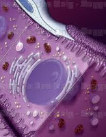
Hepatocytes are the most numerous cells of the liver, represent 80% of the volume and about 60% by number. Their shape is multifaceted, with a number of areas ranging from six to twelve, their diameter ranges from 20 to 30 microns. They are often
multinucleated and
tetraploid, with the number of cores that can be up to four, a large nucleolus, rough and smooth endoplasmic reticulum well developed, numerous
cisternae of
Golgi, ribosomes, lysosomes, mitochondria, peroxisomes, which are both one of the cell types in which the organelles are more developed and presented, due to the high metabolic needs and the wide variety of tasks which they perform.
In a well-nourished body is not difficult to detect moderate amounts of glycogen and lipid vacuoles, or in the case of an overdose of iron, vacuoles or aggregates of ferritin and hemosiderin. The cytoplasm is eosinophilic background for the large number of mitochondria but with numerous basophilic granules due to the rough
endoplasmic reticulum and ribosomes. You can find lipofuscin granules of golden-brown. The pole is fitted with sine
dell epatocita many long and
irregular microvilli on average 0.5 microns, the surface area alone is equal to 2 / 3 of the entire cell. Two adjacent hepatocytes form their plasma membranes with the
bile canaliculi and are joined by tight junctions to prevent the entry of bile into the interstices, in the rest of the cell are the most common desmosomes and gap junctions. At the level of bile canaliculi accumulating numerous exocytosis of vesicles containing precisely to secrete bile into canaliculi.
Liver Cells
Ito or
stellate cells, mesenchymal and far fewer of
hepatocytes, are placed between the plates at the base of
hepatocytes, and have a star-shaped or irregular. Their
cytoplasm is rich in
lipid vesicles containing vitamin A, and their job is to secrete the main constituent materials of the matrix, including
type III collagen and
reticulin. They are essential in liver regeneration following injury or surgery because they secrete growth factors responsible for good regeneration capacity of the liver. In case of injury can replace damaged hepatocytes and by the
secretion of collagen and other structural proteins, forming scar tissue from the area 3 of each berry. Other substances secreted from their body contribute to
homeostasis.
Liver Cells
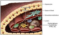
Sinusoidal endothelial cells are
fenestrated endothelium of
venous sinusoids of the
liver. They have flattened, with an oval centrally located, scant cytoplasm containing numerous vesicles
transcitotiche, are joined by adherens junctions. The perforation between these cells are very large and combined in complex with an average diameter of 100 microns, so that blood can easily spill over the
space of Disse and come into contact with the
microvilli of hepatocytes.
Liver Cells
Kupffer cells, macrophages of the
liver, are derived
monocytes, they are in the lumen of the venous sinusoids. Their shape is variable and irregular, with numerous
protrusions typical of cells of the macrophage that extend into the lumen of the sinusoid. Their function is to remove any debris by
phagocytosis in the blood flow in the liver, but may also stimulate the immune system through the secretion of numerous factors and
cytokines. Remove aged or damaged red blood cells acting as a complement to the spleen (which may be substituted in case of
splenectomy).
Liver Cells


No comments:
Post a Comment